Lengthening Anterior Teeth With Composite

Terry Shaw

General Dentist

Perth-Ando​ver, N.B, Ca​nada

7,622 views

Introduction to the case

69 year old gentleman came in for consult on bonding. Recent crown on right canine had broken off and he was balancing it in place. Lots of wear on remaining anterior teeth. He came back 2 weeks later for an elective root canal so I could place a prefab metal post and bond his crown in place. He was back a month later when I bonded his 5 remaining anterior teeth.

Some one placed a case asking for help with lengthening anterior teeth and I discovered these pictures buried in my computer so thought I’d post them to maybe help answer some questions about the problem.

(more…)

Anterior Composite Buildup Using Bioclear Matrices

Introduction to the case

This is my 2nd Bioclear case, just looking for feedback again from all you bondadontists. This one is more Doc Terry style.

91 y.o. male, didn’t like how worn his teeth were.

Filtek SU and Beautifil flow+ again.

It’s a lot of composite, but at age 91 I may have time on my side. You can’t tell from the pictures but the “crowns” are very thick, which I hope will compensate for the lack of posterior occlusion.

Still figuring out the technique. Big thanks to Doc Terry for making it seem possible, in the past I would have told him he needed RCT/post/crowns or a denture. This seems like a good compromise.

(more…)

Creating a Color Blend for Class V’s

4,808 views

Introduction to the case

Creating a color blend for Class V’s can be frustrating. Give this technique a try. As an added bonus you will realize a stronger bonded restoration.

Using SureFil SDR flow as the first layer over the entire surface of the prep creates a low stress strong bond to dentin. The SureFil also creates a semi translucent layer which is covered by the Filtek Supreme Ultra “Body” layer. The result is a blended composite that reflects light much like the surrounding tooth structure. If the color is off, the result will still look respectable.

(more…)

Quick Tip for Composite Symmetry

Introduction to the case

Have been asked about some tips for direct composite, especially on how to get symmetrical central incisors. This is a simple tip, but I rarely see it being done. Heck, it took me a long time to implement it. Here is how I messed up, and hopefully learned from it.

(more…)

Lateral Incisor Implant Placement and Provisional

Introduction to the case

Pt. presented to my office w/ multiple congenitally missing teeth and existing upper/lower acrylic partials. She finally made up her mind to move into implants to improve her quality of life. For #7, a Nobel Replace select 3.5 X 11.5 was placed and immediately temped w/ a screw retained provisional made over a plastic engaging cylinder w/ access hole through the cingulum. Tooth was taken out of occlusion and all excursions. Pontic was easily cut off her existing partial. I placed the top of the implant 3mm apical to where I wanted the margin of the facial tissue to settle, which buried the implant .5-1mm subcrestal. A final radiograph was taken w/ the screw retained temp in place and I noticed that the cylinder was not completely seated, probably because of the subcrestal placement. Being this is a provisional restoration, I didn’t think much of it, but was curious to see everyone’s thoughts.

When I made the provisional, I screwed down the engaging cylinder, but did not take a confirmation radiograph (mistake?). I then loosened the screw so I could pull the cylinder out with the luxatemp synch impression (mistake?). Either one of these reasons could be why the cylinder was not seating all the way on the final X-ray. Would like to hear everyone’s thoughts and comments about how they make this type of screw down provisional and if they would be concerned that it is not seated all the way. Thanks

(more…)

Replacing an Over-Contoured Crown: Lessons Learned

Introduction to the case

Replaced a failing, over-contoured crown. Was overall happy, that the case is holding up and the patient has maintained their perio regime (for the most part).

Lessons learned:

  • 1. Use of resin based temp cement especially with thin bis-acryl temporaries.

  • 2. Better communication with the lab. I had only sent them one pic. Now I send a lot more info to the lab. And most importantly…

  • 3. Minimize the prep to maximize enamel, especially with a non-discolored stump and when using lithium disilicate (and a bonding protocol for cementation).

(more…)

Single Stage Implant – Extraction, Placement, Grafting, and Loading of the Implant

Bill Schaeffer

BDS MBBS FDS RCS MRCS

The Implant Centre

4,482 views

Introduction to the case

This patient has almost perfect teeth, but then developed external resorption of their upper right central incisor. They saw an endodontist who told the patient that it wasn’t restorable, and then the patient found their way to me. The patient has kindly allowed me to show their case.

(more…)

Socket Shield (SS) Technique for Bone Preservation in Anterior Implants

872 views

Introduction to the case

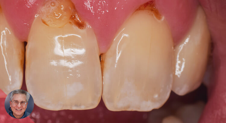
Replacing a single anterior tooth with an implant is a challenge for every dentist. The risk of losing vestibular bone height and soft tissue is unacceptable from an aesthetic point of view. The presence of osteoclasts on the inner surface of the socket walls indicates that the bundle bone will undergo resorption.

Anatomically, the buccal bone of the teeth is thinner than lingual or palatal bone. Therefore, as bundle bone is a tooth-dependent tissue, it will gradually reduce after extraction. Since there is more bundle bone in the crest of the buccal wall than the lingual wall, hard tissue loss will become most pronounced in the buccal wall (Lindhe, Clinical Periodontology and Implant Dentistry, 2008).

These scientific evidences and the clinical experience of immediate implant placement in fresh extraction sockets have led us to think that by preserving the periodontal tissues on the buccal part of the socket, we could prevent bone resorption in this critical area.

The socket-shield (SS) technique provides a promising treatment, better manages the risks, and preserves the post-extraction tissues in aesthetically challenging cases. We need to preserve and use the bundle bone to our advantage.

The principle is to prepare the root of a tooth indicated for extraction in such a manner that the buccal root section remains in-situ with its physiologic relation to the buccal plate intact. The tooth root section’s periodontal attachment (periodontal ligament (PDL), attachment fibers, vascularization, root cementum, bundle bone, alveolar bone) is intended to remain vital and undamaged so as to prevent the expected post-extraction socket remodeling and to support the buccal / facial tissues. (Howard Gluckman, Jonathan Du Toit, Maurice Salama).

(more…)